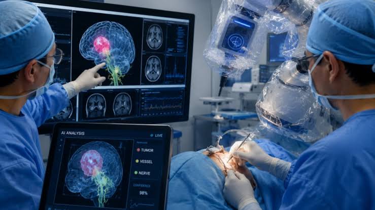

Neurosurgeons in London have used a computer-vision artificial intelligence system to guide a live brain operation, marking a major step towards the use of AI in operating rooms.
The procedure was carried out at the National Hospital for Neurology and Neurosurgery as part of a clinical trial, with software developed at University College London (UCL) analysing live video from the operation.
The AI system identified critical anatomy in real time as surgeons removed an 11mm pituitary tumour from 48-year-old Rhys Hibbert, whose worsening condition had put his eyesight at risk.
Rather than relying only on scans taken before surgery, the system processed live endoscopic footage and highlighted structures including nerves and blood vessels that surgeons needed to avoid. The surgical team retained full control of the procedure.
The technology was developed by researchers at UCL’s Hawkes Institute and trained using hundreds of annotated videos from previous endoscopic pituitary operations. It was designed to recognise critical anatomy, surgical instruments and interactions between instruments and tissue during an operation.
Pituitary surgery is particularly delicate because the tumour can be located close to the optic nerves and major blood vessels. A small error can potentially result in serious complications, including blindness or stroke.
Following the operation, Hibbert’s eyesight improved significantly and he was able to walk independently within a week, according to reports.
Researchers say the technology could eventually provide surgeons with an additional layer of real-time information during complex procedures, although further trials will be needed before wider clinical use.
The trial was funded by the UK National Institute for Health and Care Research (NIHR) and Google.